


Neonatal enteric pathogens are present on virtually all cattle farms, and neonatal diarrhea (ND) remains one of the most common diseases affecting young calves. On average, approximately one farm out of three experiences diarrhea outbreaks, with up to 50% of calves affected. Pathogen transmission can be extremely efficient.
The consequences include increased workload, farmer distress, impaired calf welfare, reduced growth performance, disruption of gut microbiota, an increased risk of bovine respiratory disease (BRD), and, in some cases, death.
As the risk of ND is highest during the first weeks of life, close monitoring of calves during this critical period is essential. Any suspected case should be investigated promptly to limit pathogen transmission and reduce the impact on animal health and performance.
Prefer to listen to this article? Click the play button below and enjoy our podcast!
1- What are the early symptoms of neonatal diarrhea in calves?
As with many infectious diseases, particularly BRD, calves suffering from neonatal diarrhea often appear dull and depressed. Careful observation from a distance may reveal subtle changes in posture and behaviour, including a lowered head and ears, an arched back, reduced alertness, increased time spent lying down, and reduced milk intake.
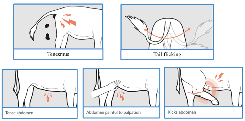
Because enteric infections are frequently associated with abdominal pain, affected calves may show signs of discomfort such as a tense facial expression, bruxism (teeth grinding), tenesmus during defecation, and frequent tail flicking. Abdominal pain may also be evidenced by kicking at the abdomen and increased sensitivity to palpation or handling.
Sensors can be valuable tools for the early detection of reduced milk intake and activity, often identifying calves at risk before obvious clinical signs become apparent.
2- To what extent is the calf affected?
A calf with neonatal diarrhea can deteriorate rapidly due to dehydration, electrolyte losses, metabolic acidosis, hypoglycemia, and, in some cases, septicemia. Early assessment of disease severity is therefore essential to ensure timely and appropriate treatment.
The clinical examination of a scouring calf should include:
- Rectal temperature measurement. Hypothermia is generally associated with a poorer prognosis than hyperthermia.
- Navel examination for signs of omphalitis.
- Abdominal examination to assess distension, fluid accumulation, or abdominal tuck.
- Assessment of distal limb temperature, as cold extremities may indicate poor peripheral perfusion.
- Evaluation of posture, gait, and ability to stand, including signs of weakness, ataxia, or a staggering gait.
- Examination of the mucous membranes (colour and moisture): pink, pale, congested, cyanotic, moist, dry, or tacky.
- Fecal evaluation.
- Assessment of dehydration status.
How is faecal consistency scored in calves with diarrhoea?
The fecal score is useful for identifying calves with diarrhea: 0 = normal (firm but not hard); 1 = soft (does not hold form, piles but spreads slightly); 2 = runny (spreads readily); and 3 = watery (liquid consistency, splatters). A score of ≥2 indicated the presence of diarrhea.
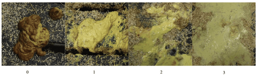
Renaud D.L. et al, 2020
From a fecal score of 2 onwards, the calf should be evaluated for dehydration, and any dehydration identified should be corrected promptly to prevent further deterioration.
How can dehydration be assessed in calves with neonatal diarrhoea?
Key indicators of dehydration include calf demeanor, eyeball recession (enophthalmos), and skin tent duration.
Guidelines for assessment of hydration status in calves with diarrhea (adapted from Smith G., 2009)
Depressed calves are lethargic, spend more time lying down, and show reduced milk intake. Comatose calves are non-responsive to their surroundings, remain recumbent, are unable to stand, and refuse to nurse.
The most reliable field method for estimating dehydration is the assessment of eyeball recession (enophthalmos) at the medial canthus. The distance (in millimetres) between the eyeball and the lower eyelid is measured. As dehydration progresses, the eye recedes further into the orbit, increasing this distance.
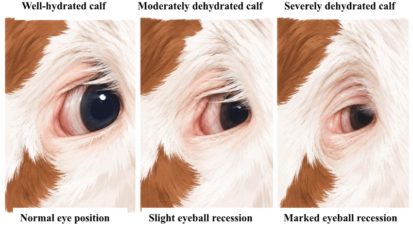
Image created with AI, adapted from Constable P.D. et al, 2021
Skin tent duration can also be used to assess hydration status. It is measured by gently pinching and lifting the skin of the neck or upper eyelid and recording the time required for the skin to return to its normal position. The longer the skin remains tented, the greater the degree of dehydration.
After assessing hydration status, neurological status and reflexes should be evaluated to identify calves suffering from metabolic acidosis.
What are the clinical signs of metabolic acidosis in calves with diarrhoea?
Key signs of metabolic acidosis are neurological and neuromuscular rather than digestive.
- Depression / dullness
- Reduced suckle reflex
- Weakness
- Ataxia ("drunken" gait)
- Increased recumbency
- Reduced menace and panniculus reflexes
- Deep, rapid breathing (compensatory hyperventilation)
- Inability to stand (severe cases)
- Coma (advanced cases)
Which clinical signs suggest septicemia in calves with neonatal diarrhoea?
Finally, the clinical examination should include an assessment for signs of septicemia, as bacteremia and septicemia may complicate neonatal diarrhea and significantly worsen the prognosis.
Suggestive signs of septicemia include:
- Hypothermia
- Cold extremities and poor peripheral perfusion
- Injected, congested, or pale mucous membranes
- Prolonged capillary refill time
- Tachycardia
- Petechiae or scleral injection
3- Which ancillary diagnostics help assess severity and guide treatment?
While the clinical examination remains the cornerstone of severity assessment, ancillary diagnostics can help quantify abnormalities and refine treatment decisions. Two calves can produce equally watery diarrhoea and require completely different treatment!
A wide range of blood parameters can now be measured on-farm using point-of-care diagnostics.
4- Which analyses are needed to identify the potential causes of the episode?
Identifying the causative agent(s) helps confirm the diagnosis, implement appropriate control measures, and assess the risk for other calves. Since mixed infections are common, testing should ideally target the main enteric pathogens simultaneously.
The main pathogens to investigate in cases of neonatal calf diarrhea include Rotavirus, Coronavirus, Cryptosporidium parvum, enterotoxigenic Escherichia coli, Salmonella spp., and Clostridium spp.
- The ideal sample is fresh feces collected directly from the rectum of 3 to 5 untreated calves during the early stage of disease. Select calves showing typical clinical signs. Avoid calves that have already received antimicrobial treatment whenever possible and avoid chronic cases or calves recovering from diarrhea. Collect fresh feces directly from the rectum. Besides rapid antigen tests, additional diagnostic methods include PCR, bacteriology, and antimicrobial susceptibility testing.
- Blood sampling is recommended when septicemia is suspected: a complete blood count (CBC), fibrinogen, and haptoglobin concentrations can be measured to assess inflammation and infection. Blood culture, when feasible, may help identify bacteremia. Measurement of serum IgG concentration or total serum protein can be used to evaluate transfer of passive immunity (TPI).
- Necropsy recently deceased calves whenever possible: Collect samples as soon as possible after death to minimize tissue autolysis and submit:
- Small intestine (jejunum and ileum), colon; intestinal contents: collect 10-30 g in a sterile container or submit a 10-15 cm ligated segment of intestine containing intestinal contents.
- Mesenteric lymph nodes
- Liver and spleen (especially if septicemia is suspected)
- Another useful investigation during a neonatal diarrhea outbreak is to assess the quality of passive immunity transfer within the herd. Both primiparous and multiparous cows should be included. Colostrum quality can be evaluated using a Brix refractometer or by measuring IgG concentration, while passive immunity transfer in calves can be assessed through serum IgG concentration or total serum protein measurement. This approach allows the identification of deficiencies in colostrum quality, colostrum management, or passive immunity transfer that may contribute to the occurrence and severity of neonatal diarrhea.
- Finally, sampling drinking water may also be relevant, particularly when waterborne pathogens are suspected or when recurrent outbreaks occur despite appropriate preventive measures. Water quality assessment should include microbiological testing (e.g., total bacterial count, coliforms, Escherichia coli) and, when indicated, evaluation of physicochemical parameters such as pH, mineral content, or contamination by organic matter. Poor water quality may contribute to pathogen transmission and increase the risk of enteric disease in young calves.
5- How should diagnostic results be interpreted?
A positive test result does not necessarily identify the cause of the outbreak. The relevance of a detected pathogen depends on its consistency with the calf's age, clinical presentation, lesion pattern, and herd epidemiology. Diagnostic results should always be interpreted within the broader clinical and management context.
Three key questions should be answered when interpreting diagnostic results:
- Is the detected pathogen consistent with the age of the affected calves?
- Is the pathogen compatible with the clinical signs and lesions observed?
- Is the pathogen consistently detected in affected calves?
Take-home messages
- Early detection relies on routine observation of fecal consistency, appetite, and behavior.
- Severity is driven primarily by dehydration and metabolic acidosis rather than fecal score alone.
- Pathogen detection must always be interpreted in the context of age, clinical signs, and epidemiology.
- Failure of passive transfer, inadequate hygiene, and high environmental pathogen pressure remain the most important risk factors.
- Successful NCD control requires combining diagnostics with a thorough assessment of management practices on the farm.
References
Ramos J.S. et al.: The burden of disease and syndromes in preweaning dairy calves at the farm level: Estimation based on the disability-adjusted life years (DALY) approach. Journal of Dairy Science, Vol. 108 No. 10:11376–11388.
Johnson, K.F.; Chancellor, N.; Wathes, D.C. A Cohort Study Risk Factor Analysis for Endemic Disease in Pre-Weaned Dairy Heifer Calves. Animals 2021, 11, 378.
Dillane P., Krump L., Kennedy E., Sayers R.G., Sayers G.P.: Determining the predictive capability of a Clinical Assessment Scoring Chart to differentiate severity of the clinical consequences of neonatal calf diarrhea relative to gold-standard blood gas analysis. 2020, PLoS ONE 15(4): e0230708.
Renaud D., Buss L., Wilms J.M., Steele M.A.: Technical note: Is fecal consistency scoring an accurate measure of fecal dry matter in dairy calves? Journal of Dairy Science, 2020; 103, 10709-10714.
Smith G.W.: Treatment of Calf Diarrhea: Oral Fluid Therapy. Vet Clin Food Anim 2009, 25: 55–72.
Constable PD, Trefz FM, Sen I, Berchtold J, Nouri M, Smith G, Grünberg W. Intravenous and Oral Fluid Therapy in Neonatal Calves With Diarrhea or Sepsis and in Adult Cattle. Front Vet Sci. 2021 Jan 27;7:603358.
Kasari T.R.: Metabolic Acidosis in Calves. Veterinary Clinics of North America: Food Animal Practice 1999, Volume 15, Issue 3, 473-486.
Trefz F.M. et al: Metabolic acidosis in neonatal calf diarrhea-clinical findings and theoretical assessment of a simple treatment protocol. J Vet Intern Med. 2012, 26(1):162-70.
Bellino C. et al: Development of a diagnostic diagram for rapid field assessment of acidosis severity in diarrheic calves. J Am Vet Med Assoc 2012; 240(3): 312-6.
Van Metre D.C., Tennant B.C., Whitlock R.H.: Infectious Diseases of the Gastrointestinal Tract. Rebhun's Diseases of Dairy Cattle. 2008:200–94.
Constable P.D. et al: Intravenous and Oral Fluid Therapy in Neonatal Calves With Diarrhea or Sepsis and in Adult Cattle. Front Vet Sci. 2021, 27;7:603358.

Leave your comments here