The Epizootic Haemorrhagic Disease (EHD) is a viral disease of wild and domestic ruminants. It was first diagnosed in North America in 1955 in a white-tailed deer. In this species, it is responsible for significant clinical signs and high mortality. In domestic ruminants (cattle, sheep, goats and camelids), its clinical expression is different and often less severe, but its recent spread to new territories is a real concern for livestock farming.

EHD is a vector-borne disease. Direct transmission from one animal to another is not possible. The disease is transmitted by a small biting midge belonging to the Culicoides genus. This characteristic explains the seasonal nature of the disease. Infections are most frequent during periods of high insect presence, i.e. late summer and autumn. However, as winters become less severe, the insect's activity can extend into the cold season. It is therefore possible, as it has been seen for the Blue-Tongue Disease, for cases to be identified in winter or spring. The same climatic reasons may also be behind the geographical spread of the disease.
It is interesting to note that EHDV belongs to the same genus as Blue-Tongue virus (BTV), and that the vector is also the same.
Clinical signs
In White-tailed deer, the peracute form is characterized by high fever, anorexia, respiratory distress and edema of the face and neck. The outcome is often fatal within a few days. In the classic (acute) form, bleeding and ulcerative lesions of the tongue, palate, gums, rumen and abomasum. In the chronic form, animals limp, feed less and die.
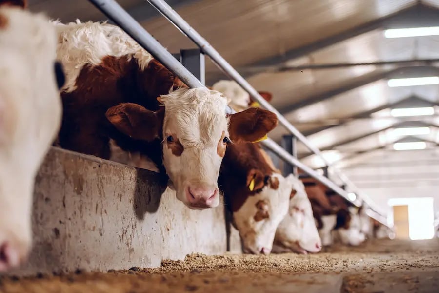
In cattle, clinical expression varies according to serotype, but is fairly similar to that caused by BTV. A strain close to EHDV serotype 2, the Ibaraki virus, seems to be the most pathogenic. Affected animals become dehydrated, lose weight and sometimes die of pneumonia. Other serotypes (1, 6, 7 and apparently 8, recently described in Europe) have a less severe clinical expression. Affected animals generally present with fever, anorexia, mouth ulcers, difficulty breathing and swallowing. Ocular discharge, erythema and lameness have also been described. Unlike White-Tailed deer, mortality in cattle is rare.
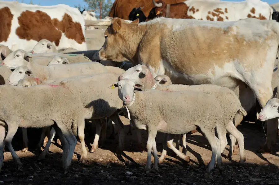
In small ruminants and camelids, the disease is sub-clinical and therefore inapparent. To date, it has not been established whether these species constitute a reservoir for the disease. But if they were, their role in the spread of the disease would be major.
Control
No specific treatment exists for Epizootic Haemorrhagic Disease.
Regarding prevention, at individual level, treatments against external parasites to prevent the risk of culicoides bites can be envisaged, but their efficacy is far from proven. On a collective scale, to limit the geographical spread of the disease, diagnosis is essential in order to determine the herds in which the virus is circulating and to take the appropriate measures. Within the European Union, the disease is regulated. Control measures are based on restrictions on animal movements from infected areas. However, according to the French Agency for Food, Environmental and Occupational Health and Safety (Anses), the effectiveness of these measures "remains low".
Vaccines have been marketed in the USA and Japan. However, due to the antigenic variability between EHBV serotypes, vaccines are only effective against the serotype they contain. As the strain currently speading in Europe belongs to another serotype (serotype 8), vaccination is not yet a solution.
Key messages
- The Epizootic Haemorrhagic Disease (EHD) is a vector-borne disease of wild and domestic ruminants.
- The virus is transmitted by a biting midge belonging to the Culicoides genus.
- Clinical signs in cattle are dominated by fever and anorexia. Mouth ulcers and respiratory difficulties are also possible. However, death is rare.
- In sheep, goats and camelids, the disease is generally sub-clinical.
- Only diagnosis and restrictions on the movement of animals from infected areas can limit the spread of the disease.

References
Zientara, S., Sailleau, C., Viarouge, C., Desprat, A., Belbis, G., & Bréard, E. (2011). La maladie hémorragiqueépizootique des cervidés succédera-t-elle à la fièvrecatarrhale ovine?.
https://www.anses.fr/fr/virus-bovins-moucherons-emerge-en-europe accessed on line 22.09.23
https://www.woah.org/fileadmin/Home/eng/Health_standards/tahm/3.01.07_EHD.pdf accessed on line 22.09.23
Leave your comments here